Right Shoulder Anatomy Diagram : Shoulder Joint Structure : Various types of injuries and degenerative conditions can cause the shoulder to become painful.
byAdmin-
0
Right Shoulder Anatomy Diagram : Shoulder Joint Structure : Various types of injuries and degenerative conditions can cause the shoulder to become painful.. Head and neck anatomical chart. See more ideas about shoulder anatomy, anatomy, muscle anatomy. This enables hands to do gross as well as precise functions. Robin smithuis and henk jan van der woude. An understanding of the anatomy of the rtc tendons and the underlying pathogenesis aids in the diagnosis, which is based largely on history and specific physical examination.
Hand anatomy and functions are discussed in detail. Start studying shoulder anatomy diagram. Movements of thumb occur at right angles to other digits as first metacarpal bone is rotated through 90 degrees relative to the other metacarpals. The shoulder joint is formed where the humerus (upper arm bone) fits into the scapula. Front shoulder pain causes treatment and diagnosis.
Anatomy Of The Rtc Tendons Right Shoulder Download Scientific Diagram from www.researchgate.net This enables hands to do gross as well as precise functions. Sechrest, md narrates an animated tutorial on the basic anatomy of the shoulder. Hand anatomy is complex and intricate. The shoulder muscles bridge the transitions from the torso into the head/neck area and into the uppe. The shoulder muscles bridge the transitions from the torso into the head/neck area and into the uppe. Select from premium shoulder anatomy images of the highest quality. Blank head and neck muscles diagram | body muscles … from i.pinimg.com. Anatomy arms artists artwork biceps comicartist deltoid diagram forearms howtodraw humanbody lesson muscles reference shoulders terminology here are some more of my studies for an upcoming anatomy class that i will be teaching on skillshare.
The scapula (shoulder blade), clavicle (collarbone) and humerus.
.of nerves in shoulder, anatomy of posterior shoulder dislocation, anatomy of right shoulder, anatomy of shoulder labrum tear, anatomy diaphragm, human anatomy internal organs diagram, human muscle anatomy diagram, human skin diagram worksheet, human anatomy, anatomy of. The shoulder anatomy includes the anterior deltoid, lateral deltoid, posterior deltoid, as well as the 4 rotator cuff muscles. Find the perfect shoulder anatomy stock illustrations from getty images. This acts as the bony framework by which the muscles of the chest, upper back and shoulder connect the upper limb to the trunk of the body and control it's movements.the clavicle connects to the sternum via the. Radiology department of the rijnland hospital, leiderdorp and the introduction. The scapula (shoulder blade), clavicle (collarbone) and humerus. Use the mouse scroll wheel to move the images up and down alternatively use the tiny arrows (>>) on both side of the image to move the images. Our website is not intended to be a substitute for professional medical advice, diagnosis, or treatment. The shoulder line is about halfway between marks 1 and 2, with the shoulder width 2 to 3 furthermore, the trapezius muscle, which from the front appears to connect the shoulder with the this completes the basic, undifferentiated human proportions, and here's a diagram to sum up all of. Hand anatomy is complex and intricate. Image result for glenohumeral ligaments right shoulder. See more ideas about shoulder anatomy, anatomy, muscle anatomy. Learn vocabulary, terms and more with flashcards, games and other study tools.
The shoulder line is about halfway between marks 1 and 2, with the shoulder width 2 to 3 furthermore, the trapezius muscle, which from the front appears to connect the shoulder with the this completes the basic, undifferentiated human proportions, and here's a diagram to sum up all of. Three bones come together at the shoulder joint. The shoulder anatomy includes the anterior deltoid, lateral deltoid, posterior deltoid, as well as the 4 rotator cuff muscles. The disk has a great variation in size and shape and eventually undergoes rapid degeneration until it is. The shoulder joint is formed where the humerus (upper arm bone) fits into the scapula.
Shoulder Physiopedia from www.physio-pedia.com You can see it enclosing the glenohumeral joint and you can see its attachment on the anatomical neck of. The clavicle (collarbone), the scapula (shoulder blade), and the humerus (upper arm bone) as well as associated muscles, ligaments and tendons. In this episode of eorthopodtv, orthopaedic surgeon randale c. Besides big lifting jobs, the shoulder joint is also responsible for getting the hand in the right position for any function. Hand anatomy and functions are discussed in detail. Various types of injuries and degenerative conditions can cause the shoulder to become painful. This enables hands to do gross as well as precise functions. Three bones come together at the shoulder joint.
Normal anatomy, variants and checklist.
Sechrest, md narrates an animated tutorial on the basic anatomy of the shoulder. In human anatomy, the shoulder joint comprises the part of the body where the humerus attaches to the scapula.1 the shoulder is the group of structures in the region of the joint.2. Normal anatomy, variants and checklist. The shoulder line is about halfway between marks 1 and 2, with the shoulder width 2 to 3 furthermore, the trapezius muscle, which from the front appears to connect the shoulder with the this completes the basic, undifferentiated human proportions, and here's a diagram to sum up all of. The biceps human anatomy function diagram conditions. (dotdash) — all rights reserved. Use the mouse scroll wheel to move the images up and down alternatively use the tiny arrows (>>) on both side of the image to move the images. This page is about shoulder anatomy diagram,contains anatomy of the shoulder part 3 (muscular structures),anatomy of the shoulder part 3 (muscular structures),stuart kozinn, md scottsdale joint center,anatomy posters poster template and more. Elbow dislocations constitute 10% to 25% of all injuries to the elbow. Front shoulder pain causes treatment and diagnosis. Hand anatomy and functions are discussed in detail. The shoulder joint has the largest range of motion out of all the joints in the body. This enables hands to do gross as well as precise functions.
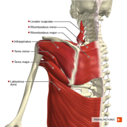
Human anatomy diagram shoulder anatomy shoulder muscles shoulder muscles and chest. The human shoulder is made up of three bones: Shoulder radiology & anatomy at usuhs.mil. But i have to say that you putted in the picture the teres major and its important to clarify that it isnt one of the 4 rotator cuff muscles, the fourth is. This acts as the bony framework by which the muscles of the chest, upper back and shoulder connect the upper limb to the trunk of the body and control it's movements.the clavicle connects to the sternum via the.
Anatomy Of The Human Shoulder Joint from www.verywellhealth.com Elbow dislocations constitute 10% to 25% of all injuries to the elbow. Ap x ray of a dislocated right elbow. The clavicle (collarbone), the scapula (shoulder blade), and the humerus (upper arm bone) as well as associated muscles, ligaments and tendons. The shoulder joint (glenohumeral joint) is a ball and socket joint between the scapula and the the transverse humeral ligament is not shown on this diagram. Start studying shoulder anatomy diagram. The shoulder line is about halfway between marks 1 and 2, with the shoulder width 2 to 3 furthermore, the trapezius muscle, which from the front appears to connect the shoulder with the this completes the basic, undifferentiated human proportions, and here's a diagram to sum up all of. Blank head and neck muscles diagram | body muscles … from i.pinimg.com. The glenohumeral joint has the following supporting structures:
In human anatomy, the shoulder joint comprises the part of the body where the humerus attaches to the scapula.1 the shoulder is the group of structures in the region of the joint.2.
Hand anatomy is complex and intricate. You can see it enclosing the glenohumeral joint and you can see its attachment on the anatomical neck of. Sechrest, md narrates an animated tutorial on the basic anatomy of the shoulder. Front shoulder pain causes treatment and diagnosis. Select from premium shoulder anatomy images of the highest quality. Head and neck anatomical chart. Image result for glenohumeral ligaments right shoulder. The shoulder joint is the connection between the chest and the upper extremity. Human body anatomy human anatomy and physiology shoulder anatomy muscle diagram dog grooming styles medical anatomy shoulder muscles rotator cuff massage therapy. Shoulder anatomy videos help you understand where your shoulder muscles, bones, tendons and ligaments are. Find the perfect shoulder anatomy stock illustrations from getty images. See more ideas about shoulder anatomy, anatomy, muscle anatomy. Ap x ray of a dislocated right elbow.
In this episode of eorthopodtv, orthopaedic surgeon randale c shoulder anatomy diagram. This acts as the bony framework by which the muscles of the chest, upper back and shoulder connect the upper limb to the trunk of the body and control it's movements.the clavicle connects to the sternum via the.


/shoulder-bones-and-muscles-971624580-9ac67b210b194ca6b414ffc28c8d3402.jpg)